


일자: 2016년 9월 2일(금)
연자: Dr. Filippo Piccinini, Ph.D
Advanced Research Center on Electronic Systems (ARCES), University of Bologna
Title:
Quantitative microscopy using 3D multicellular spheroids: generation, imaging, and analysis.
Summary of the presentation:
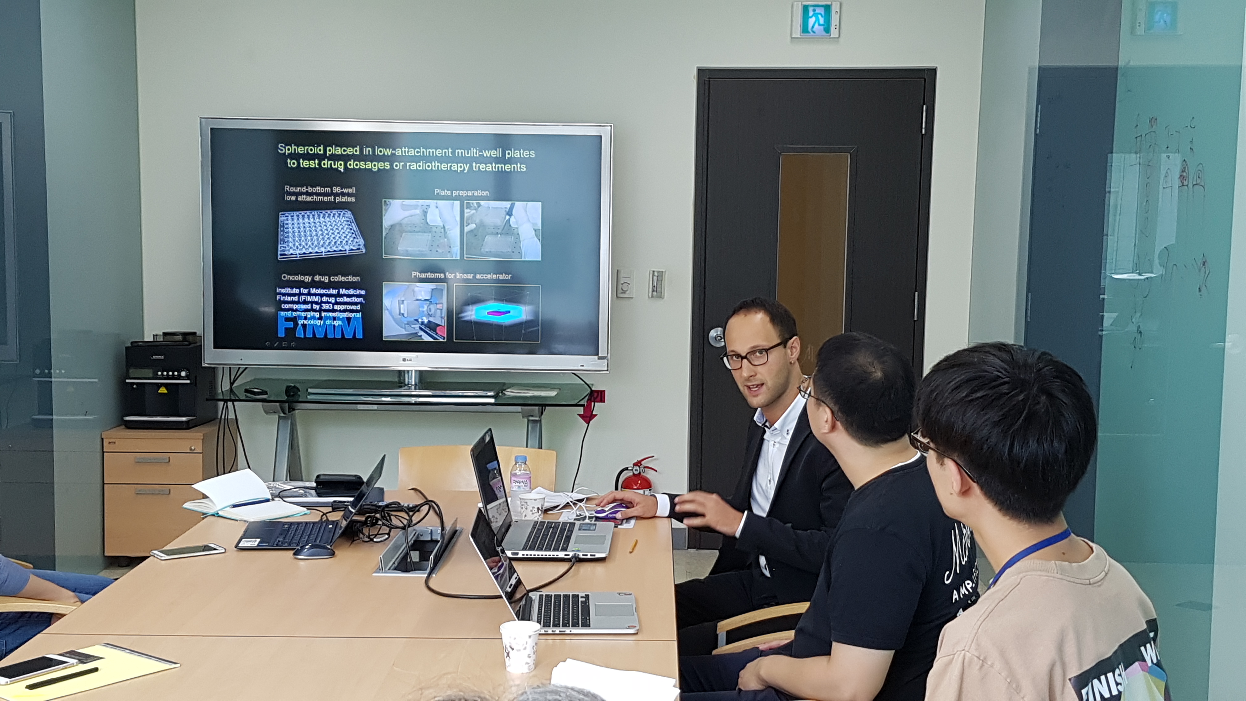
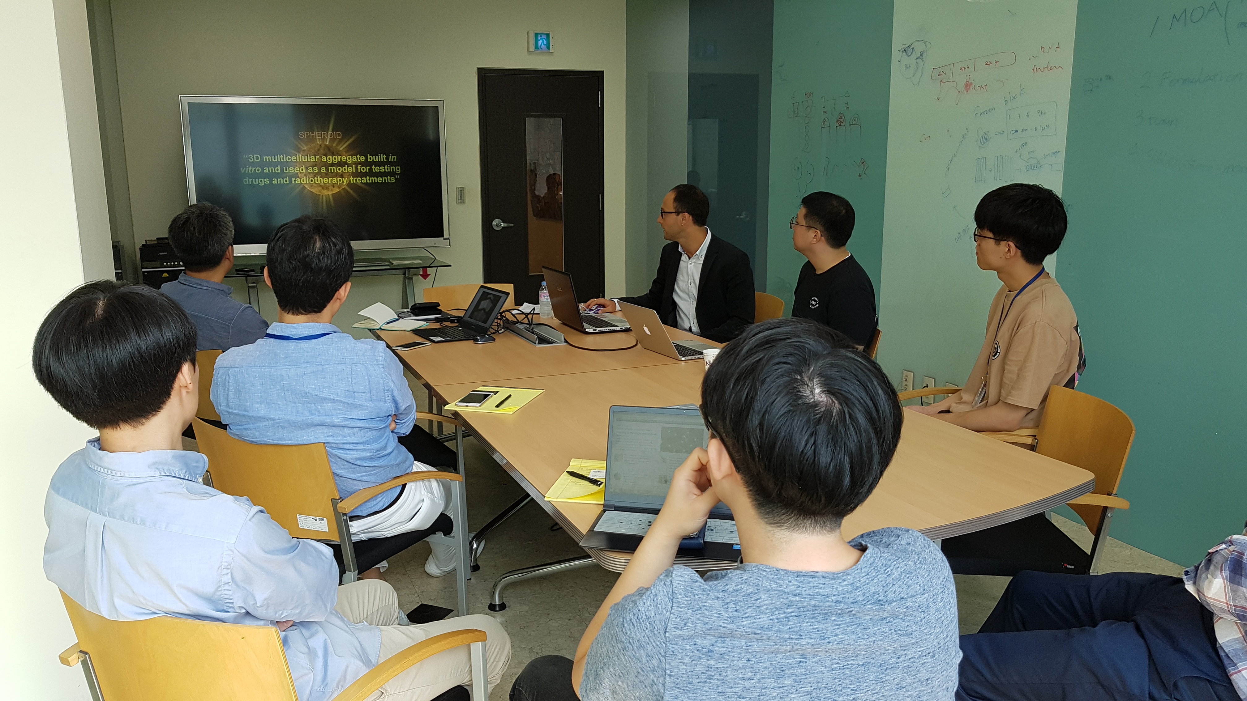
Three-dimensional (3D) multicellular aggregates, typically known as spheroids, are in vitro models widely used for testing drugs and radiotherapy treatments. However, three main open-problems jeopardize the data reproducibility when using 3D spheroids as in vitro models: (a) homogeneity and stability of the generated spheroids; (b) penetration depth of the microscopes used for imaging the spheroids; (c) lack of software tools designed to analyse the spheroids. In a recent study we compared different systems available to generate multicellular spheroids. By using a light-sheet microscope we analysed over-time homogeneity and stability of the spheroids obtained, proving that a spheroid pre-selection based on a morphological analysis is needed to obtain statistical relevant data. Then, we designed AnaSP (http://sourceforge.net/p/anasp/) and ReViSP (http://sourceforge.net/p/revisp/), freely available software tools to segment and extract morphological features of the spheroids (e.g. volume), to guide researchers in performing experiments based on 3D models. Finally, we developed CellTracker (www.celltracker.website) and Advanced Cell Classifier (www.cellclassifier.org), open-source software tools for performing cell tracking and classification, today using 2D cell cultures but soon extended to 3D analyses.